Russian English
Russian English
INTRODUCTION Point of view
METHOD
POSSIBILITIES of METHOD
PROGNOSIS
for SPONSORS and INVESTORS
HISTORY
THEORY of the METHOD
RESULTS
CONDITIONS
e-mail: Write to us
Our awards
Rambler's Top100
Rambler's Top100


 www.guestbook.ru - «ãç訩 á¥à¢¥à £®á⥢ëå ª­¨£
Rehabilitation of moving activity and nervous-muscular coordination at defeats of the central nervous system after Skorbun-Zverev method: removal of spasticity, atonic muscles tone restoration, accelerated record of base movements in a memory 

Results
In this section the frames from films and photos is given with which we have tried to illustrate productivity of a method. Usually shootings were conducted at the beginning of a course, and then before the next training.
TANJA, 4 years old. Diplegia

09.09.98 Shooting prior to the beginning of rehabilitation course.


Disability, atony of muscles of a back does not allow to keep a pose.

It is all that can make Tanja in attempt to put a leg on a leg

The muscles of legs are disabled. Foots (ankle joint) are so weakened that she can not stand and go without footwear. Even after five trainings the foot is so weakened, that it is possible to turn it without a pain almost on 90 degrees, though the resistance is already felt.
The frames from film shooting during a course is shown below. The frames without date is shooted before the 6-th training


In spite of the fact that only some days have passed, from this frames the cardinal changes in a tone of muscles and moving activity are visible. The serviceability of muscles of a back, legs is restored. Now Tanja not only can normally to sit and to stand, but also to pass from a support to a support independently, and even shifts its toys. On the bottom staff Tanja being without footwear.

...................................................................

FRANSOIS O. Spinal brain injury
See
Spinal brain injury

...................................................................

ANDREW, 21 years old, paresis Erba, (birth trauma)

The shootings are executed during the 2-nd course. In the left column the frame is indicated, on which Andrew shows, what condition had his hand before trainings. In right one - at the moment of shootings


After therapy the wrist is passively removed in a position, close to norm

The mobility in shoulder-joint is restored in complete volume
Can chin on two hands, that it was impossible earlier. Before contractures of a wrist and elbow joints simply did not allow to make a handfast of a bar

According to Anrew's words, the sensation has appeared that pectoral muscles and latissimus dorsi muscles have begun to grow.
Our first patient with Cerebral Palsy
ALEKSANDRA S. (was born October 1994).

First series of stimulations carried out in the second half of December 1996). After that there was 10 courses of VS-treatment. In 1999 the diagnosis CP was removed


An extract from the case record. (12 December 1997. Doctor-orthopedist-..)

...In the early neonatal period had natal asphyxia, subarachnoid hemorrhage; purulent meningitis, double pneumonia: Dysplasia of the hip joints.
Received treatment in MODOKh: to 6 months -- spacer splint, to 1 year - Vilensky splint. Was given massage, thermal treatment, electrophoresis with Ca, P.

Under observation by local neurologist.
Diagnosis: Perinatal encephalopathy, period of rehabilitation. 
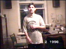
Infantile cerebral paralysis. Hemiparesis at the left of spastic type, more evident in lower limb.

Received 3 series of treatment in NTTs PNI (Skvortsov-Osipenko method).

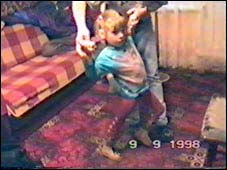
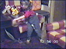
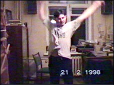
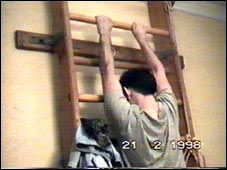
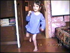
Began to independently walk at age of 1 year 2 months. Gait changed. Pronounced ataxia. "Drags" left leg. Movements in left hip joint limited - bends to 50 degrees, swing to 30 degrees. Left hip shortened by 1 cm.
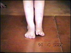
Active movements not possible in left ankle joint, passive - very limited. Equinovarus. Umbilical hernia.

Contracture of left hip joint owing to innate dislocation of left hip. Aseptic necrosis of the head of the left hip, repair stage. Contracture of left ankle joint of neurologic genesis.

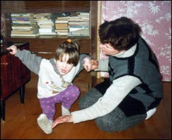
Received PT, massage, removable splint (for night) for correcting left ankle joint.

Operated on in the city of Tula in the IKR according to V.B. Ulzibat method on 9 June 1997. Multiple fibrositis.

Mptomography of brain 11 November 1995.
-- focal lesion of brain according to demyelinization type process, hypoplasia of the corpus callosum. 1996
EEG - changes in brain showing rough stimulation of the truncal structures of the brain, formation of an epiactive focus in deep structures of the brain. (before stimulation)
EMG - suprasegmental character of damage. Speed of carrying out pulse by efferents in N. Tendinous reflexes S=D, caused.
Consultation with assistant Professor Shariborova - thought should be given to myelodysplasia at the level of the lumbar section of the spinal cord.

after VS-therapy
EEG of 17 September 1997 -- specific features of epiactivity not discerned.

Examination by neurologist 27 October 1997 (after 3 coursis of treatment by VS-therapy)
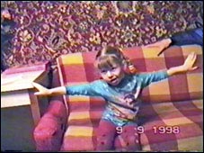
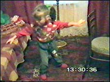
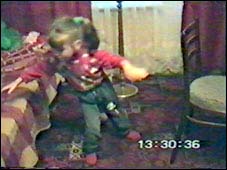
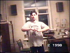
Began to move more freely. Symptoms of ataxia less evident. Sparing gait, drags left leg, left foot turned inwards. Tonus of left limbs high, greater in leg. Reflexes S>D. Speech defects. Girl communicative, quiet.
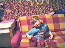
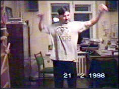
Examination by orthopedist 10 November 1997
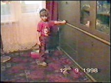
Gait became freer. During walking steps on whole foot. Varus setting of left foot. 
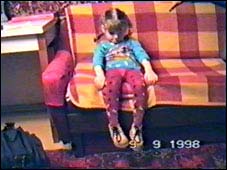
Mobility of right hip joint complete, left limited: - swinging to 45 degrees, bending to 150 degrees. Mobility in knee joints complete. Right ankle joint and foot - N. Left ankle joint - independent movements within the limits of 5 degrees, passive movements limited. The foot is passively put in the correct position...

Under observation of mother (summer 1997):
I began to note that the toes of the left foot hardly moved, even at age less than one year. But left leg was like the right in respect of circumference and length.
Treatment in Skvortsov Clinic 3 times with Skvortsov-Osipenko method (Center of Neurologic Infantile Invalidism, Professor Skvortsov). After treatment the mobility (relaxation) of the hip, ankle and knee joints improved, but after 1-2 months the mobility of the joints worsened (returned to the initial state) in spite of scleromere massage (4-5 times each day), general massage (2-3 times a year). Coordination improved.
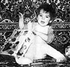
2 years old
Retarded development of left leg. Circumference of calf of left leg much less than that of right, leg has a bluish tone. Left leg spastic.

Treatment by method S.-Z. (December 1996) - 7 days. Result - mobility of ankle and hip joints increased, contracture of knee joint ended. Knee joint normal - full mobility up to present day.
After a month the condition of the ankle and hip joints slightly worsened, but as compared with condition prior to VS-treatment, the joints and muscles substantially more relaxed.

From 2 to 2.5 years old - child did not receive any treatment (on the other hand this is good, the child got a rest). The condition of the hip joint and especially the ankle joint worsened, but anyway was substantially better than prior to BMS.

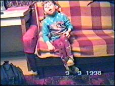
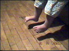
2.5 years old. Operation in Tula. After the operation the left foot takes on an almost correct (more level) position, the mobility of the hip joint substantially increases (the leg is raised to the head), but when walking the child drags his leg substantially (does not bend the knee) and walks as a whole in a much worse manner than before the operation. Furthermore, sensitivity of the sole of the left foot is in practice lost, there is no reaction to touching and tickling, cannot stir the foot, cannot stir the toes of the left foot. Cannot sit in a Turkish manner. The foot is stressed, when raised there is an abrupt stop. Free movement of the foot only inwards.


If up to therapy on our method the active movements in left ankle were impossible, and passive - hardly are limited, now passive in complete volume, and active, though have restrictions, but come nearer to normal. On a right snapshot it is visible, that the active movement of foot is possible even a range close to limiting.

Foot is restored enough, that Aleksandra could keep balance, to execute sitting, to jump on left leg


13. Discussion of results
We have shown only some examples, that illustrate rehabilitation of motor activity of the people with typical symptoms at CNS defeats (peripheral nerves only anatomically are allocated in separate system. Functionally they have not an independent significance.) - paresis, spastisity and atony. These examples proving an opportunity of re-informing of analitical departments of CNS and restoration of their work in a genetically given mode, do not show, unfortunately, that part of rehabilitation, which does not occur by jump, but requires rather long period.
Once again we shall repeat for many persons, who would like, by drinking a tablet, as in a fairy tale to find in one moment the movements, which are considered as norm. Our experience shows, that it is impossible. The removal of spasticity or setting into gear the muscles at atony is only pioneering stage, though both main and easiest. More difficult is subsequent aidless work. In memory it is necessary "to write down" images of all trajectories and conditions of a body, which then can be used at fulfilment of movement automatically.
And for recording in a memory the frequentative realized fulfilment of the appropriate movements is necessary. The process of training is easier at atony, it is more difficult at hyperkinesis, because in a memory there are stable abnormal moving stereotypes. At spastisity the process is complicated by that in a constant stress can be only òîíè÷åñêèå, slow muscle fibres. Accordingly, due to inactivity atrophy of fast moving units occurs, which provide the large force and speed. In all cases the conscientious intensive trainings are required, and on such self-restriction many persons are not capable. Nevertheless we are sure, that returning the person if not to norm than close to norm in most cases is possible.